


Heart Regeneration Research in Nonhuman Primates: A Model for Ventricular Pressure Overload
Congenital heart defects are the most common type of birth defect in the United States and can result in heart failure early in life. Stem cell grafts offer potential as a treatment for cardiac defects, but both laboratory bench experiments and whole-animal studies are necessary for progress in this area. By testing cardiac treatments in real-life situations, researchers can observe how the heart adapts to ongoing changes during exertion, such as beating more rapidly when an animal is running or climbing. ORIP’s National Primate Research Centers (NPRCs) are facilitating studies on this topic by providing expertise and resources for laboratory research, as well as new nonhuman primate (NHP) models for studying heart function in vivo.
Stem cells and regenerative medicine—the process of creating living, functional tissues to repair or replace damaged cells, tissues, or organs to treat or cure conditions caused by aging, disease, or congenital defects—are a key focus of ORIP’s research investments. A team of researchers has reported that heart muscle cells grown from human induced pluripotent stem cells can integrate into the hearts of rhesus macaques (Macaca mulatta) with a state of ventricular pressure overload, similar to what people with heart defects experience.1 This team is led by Dr. Marina Emborg, Professor, Medical Physics, and Director, Preclinical Parkinson’s Research Program, Wisconsin NPRC, University of Wisconsin–Madison (Figure 1); Dr. Jodi Scholz, Assistant Professor, Comparative Medicine, Mayo Clinic (Figure 2); and Dr. Timothy Nelson, Associate Professor, Pharmacology, Medicine, and Director, Todd and Karen Wanek Family Program for Hypoplastic Left Heart Syndrome, Mayo Clinic (Figure 3).

Dr. Emborg explained that although her research focuses on neurodegenerative disorders, her passion for this work was sparked by the death of her nephew, who had a congenital cardiac disorder. When Dr. Nelson contacted her to discuss the stem cell approach, she realized she had the opportunity to improve the lives of children born with such disorders. “If I can do something to help children with this condition, I will try,” Dr. Emborg declared. The Wisconsin NPRC is equipped with state-of-the-art facilities and skilled surgical staff who are well positioned to perform these studies in NHPs. Dr. Emborg explained that NHPs are excellent models for human disease research because of their genetic, anatomical, physiological, and behavioral similarities to humans. These animals have played key roles in understanding and treating a variety of chronic diseases.
Dr. Nelson has dedicated his career to finding a cure for congenital heart defects, using heart cells derived from the patients’ own stem cells as a treatment for children with such defects. His work has shown that pluripotent stem cell models effectively mimic biological mechanisms related to congenital heart defects, but these models are two-dimensional and lack the spatial context of the human heart.2 As an expert in NHP research, Dr. Emborg helped the team bridge this knowledge gap. Furthermore, Dr. Scholz, a trained veterinarian, has extensive experience working with various types of research animals, including NHPs.
Dr. Emborg and her team previously developed an NHP model for heart defects triggered by Parkinson’s disease. In addition to affecting movement, Parkinson’s disease can also lead to heart arrhythmias and chest pains. Dr. Emborg’s team previously studied autologous stem cells in an NHP model,3,4 which advanced to a clinical trial, Autologous-derived Study of a Parkinson’s Investigational Regenerative therapy in an Open-label trial (ASPIRO). Dr. Nelson and his colleagues were inspired by these findings and chose to apply Dr. Nelson’s approach to evaluate an NHP model for congenital heart disorders.
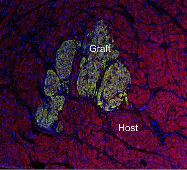
Drs. Emborg, Scholz, and Nelson tested grafts of stem cell–derived cardiomyocytes as a possible complementary treatment to traditional surgical repair of cardiac defects. Their goal was to support ventricular function and promote overall healing. The team observed that the cells integrated successfully into the muscular layer of the heart (i.e., the myocardium), matching the pattern of organization of the host cells (Figure 4). This study demonstrated the feasibility and safety of using stem cells in the first NHP model for right ventricular pressure overload, a type of cardiac problem that occurs with congenital heart disease. The authors showed that stem cells seamlessly incorporated into the muscle that is affected by the extra pressure associated with congenital heart disease. This finding paves the way for an adjunct treatment in patients to improve heart function by supplying new cardiac cells from the patient’s own stem cells.
Dr. Emborg emphasized that this NHP model is highly complex and requires expertise in numerous areas, including stem cells, surgery, and animal care. “You need a team of people that really look after the animals like they were humans. They are their patients,” she stated. Dr. Emborg explained that the Wisconsin NPRC is home to well-established expertise in stem cell biology. Dr. James Thomson, former Chief Pathologist at the Wisconsin NPRC, who is one of the pioneers of stem cell research, developed this approach in embryonic cells derived from NHPs.5 As another example of basic science being translated into a clinical setting, Dr. Thomson began performing this procedure in human cells.
As the next step in their work, the Mayo Clinic researchers are planning a clinical trial to test this approach in human patients with congenital heart defects. Dr. Emborg underscored the importance of the NPRCs for bringing together experts and enabling research that requires the use of NHPs. “Without the primate centers, this research would not be possible,” she emphasized. “You need the veterinarians who are dedicated to working with primates. There are so many key things that are needed that ORIP supports.”
ORIP is helping investigators seamlessly integrate research that can take place in a cell culture dish with whole-animal approaches. Animal models are a vital tool for testing treatments in the more realistic context of an organ embedded within a complex body system, as well as an animal interacting dynamically with its environment. Without these studies, dangerous, unanticipated consequences can occur, especially with such serious conditions as heart defects in medically fragile patients. For additional information on the ORIP-funded NPRCs and NHP resources, visit the National Primate Research Centers Consortium webpage. The ORIP Stem Cells and Regenerative Medicine webpage has more information about this research area.
References
1 Scholz J, Secreto FJ, Wobig J, et al. Human stem cell–derived cardiomyocytes integrate into the heart of monkeys with right ventricular pressure overload. Cell Transplant. 2024 Jan–Dec;33. doi:10.1177/09636897241290367.
2 Odogwu NM, Hagen C, Nelson TJ. Transcriptome studies of congenital heart diseases: identifying current gaps and therapeutic frontiers. Front Genet. 2023 Dec 13:14:1278747. doi:10.3389/fgene.2023.1278747.
3 Tao Y, Vermilyea SC, Zammit M, et al. Autologous transplant therapy alleviates motor and depressive behaviors in parkinsonian monkeys. Nat Med. 2021;27(4):632–639. doi:10.1038/s41591-021-01257-1.
4 Emborg ME, Mancinelli A, Colwell JC, et al. Preclinical evaluation of transaxial intraputaminal trajectory for enhanced distribution of grafted cells in Parkinson’s disease. J Neurosurg. 2024 Jul 26;141(6):1554–1566. doi:10.3171/2024.4.JNS24367.
5 Thomson JA, Kalishman J, Golos TG, et al. Isolation of a primate embryonic stem cell line. Proc Natl Acad Sci U S A. 1995;92(17):7844–7848. doi:10.1073/pnas.92.17.7844.