





Seeing the Invisible: Next-Generation Imaging Instruments Transform Brain Cancer Research
Brain cancer remains difficult to treat for several key reasons, one of which is the inability of drugs to successfully cross the blood–brain barrier (BBB) and modify the immunosuppressive tumor microenvironment (TME). The TME is composed of cells and vessels surrounding the tumor that provide nutrients and limit white blood cells from attacking the tumor. At least partially because of this, patients with glioblastoma (GBM), an aggressive brain cancer, have a median survival of approximately 14 months after their diagnosis,1 even with surgery, radiation, and chemotherapy. To create and advance therapies that will successfully treat brain cancer, cutting-edge imaging instrumentation is needed to capture the changing TME in real time. Imaging instrumentation allows noninvasive in vivo (in the body) capture of data for preclinical and clinical studies. In addition, imaging instrumentation provides quantifiable data for clinicians to evaluate patient progress over the course of treatment—allowing them to modify the treatment routines as needed and highlighting a key step toward personalized therapy.
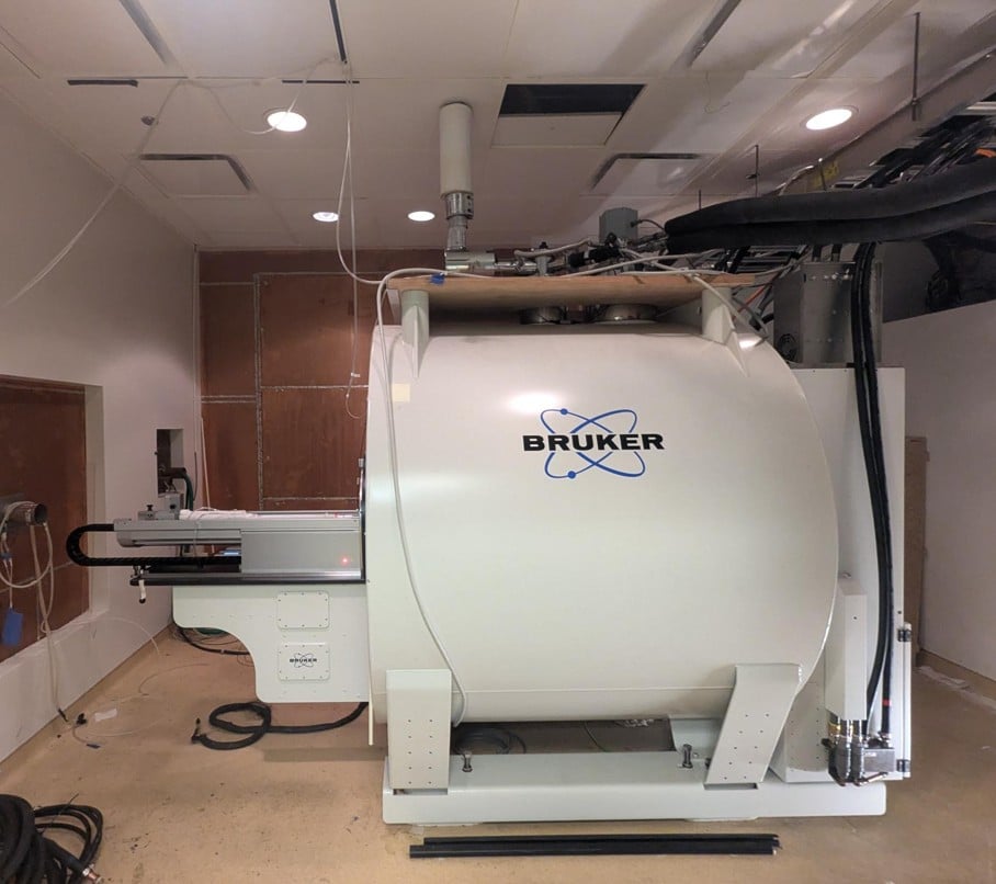
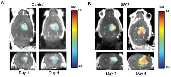
Dr. Amy B. Heimberger, Professor of Neurological Research and Jean Malnati Miller Professor of Brain Tumor Research, and Vice Chair for Research in the Department of Neurological Surgery at Northwestern Medicine (NM) Feinberg School of Medicine, is focused on understanding the underlying mechanisms of tumor-mediated immunosuppression to identify potential GBM targets for novel therapies. With funding from the National Cancer Institute (NCI) (P50CA221747), Dr. Heimberger is studying strategies that could reprogram immune cells to have antitumor traits. Through a stimulator of interferon genes (STING) pathway, the immune system senses cell stress and is activated when specific molecules are shed from dying cells. When the STING pathway is activated, tumor-associated myeloid cells can undergo changes in physical characteristics to promote activity against the tumor. Dr. Heimberger’s team has developed a potent STING agonist, IACS-8803, which, in combination with radiation therapy, increased long-term survival in a preclinical model for GBM.2 Results also showed that the combination therapy promoted BBB permeability and antitumor immune reactivity.2 “IACS-8803 transiently opened the BBB for up to 24 hours, creating a therapeutic window during which other drugs could access the tumor with far greater efficiency,” explained Dr. Heimberger. This study relied on a key piece of instrumentation—the 7-Tesla (T) Bruker Biospec 70/30 magnetic resonance imaging (MRI) scanner—from the Center for Translational Imaging (CTI) at NM Feinberg School of Medicine (Figure 1). The MRI scanner allowed Dr. Heimberger’s team to conduct accurate tumor segmentation and analyze altered anatomical intratumoral features—including size, form, and structure—in the preclinical models for GBM (Figure 2).
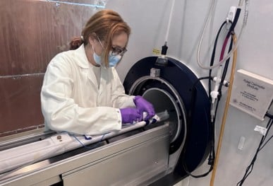
CTI is a state-of-the-art core facility for preclinical and clinical imaging instrumentation. CTI is a 9,000-square-foot facility that provides cardiology, neurology, and oncology researchers with access to several innovative imaging instruments—including a whole-body Siemens 1.5T area MRI scanner, two whole-body Siemens 3T Prisma scanners, an angiographic C-arm, and a Mediso NanoScan positron emission tomography (PET)/computed tomography (CT) scanner. Acquisition of the advanced 7T Bruker Biospec 70/30 MRI scanner in 2023 was made possible by an ORIP grant (S10OD032221) awarded to the medical school, and the MRI scanner allows CTI to support additional cutting-edge studies, such as Dr. Heimberger’s, to advance translational research and potential treatment strategies for patients (Figure 3).
Dr. Michael Markl, the principal investigator of the ORIP S10 award and CTI Director and Lester B. and Frances T. Knight Professor of Cardiac Imaging and Vice Chair for Research in the Department of Radiology at Northwestern University, emphasized that, “What excites me most about the BioSpec 7T/30 is that it fundamentally changes what we can ask of an image. The high-performance gradients, multi-channel RF coils, and dynamic shimming are not incremental upgrades—they open doors to experiments we simply could not run before.”
With additional funding from NCI (R01CA120813), Dr. Heimberger also is studying the underlying mechanisms of lymphopenia (a condition where the blood doesn’t have enough circulating white blood cells) and an anti-programmed cell death protein 1 therapeutic for GBM. In collaboration with Dr. Shulin Li, Scientific Director of Pediatrics and W. T. and Louise Jarrett Moran Distinguished Chair in Pediatric Oncology at The University of Texas MD Anderson Cancer Center, and with funding from the National Institute of Neurological Disorders and Stroke (NINDS) (R01NS122857), Dr. Heimberger is studying whether a novel neutralizing T-cell therapy (engineered white blood cells) that targets a protein called FGL2 is effective in preclinical models for GBM. GBM expresses high levels of FGL2. FGL2 suppresses the immune system and allows slow-growing tumors to become aggressive GBM.3 If successful, this therapy could progress to the next phase and help transform GBM treatment.
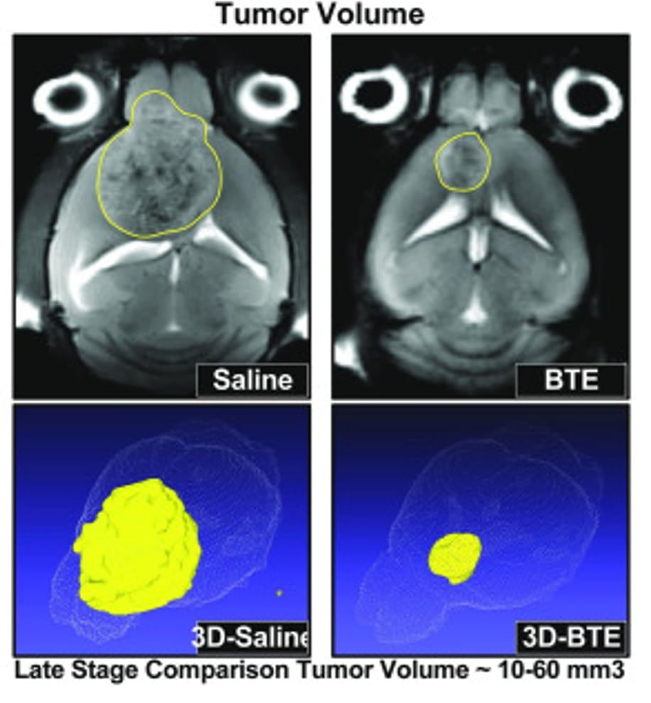 Figure 4. MRI images of tumor volume in the BTE treatment and control mice. Adapted from Zannikou et al., J Immunother Cancer (2025), licensed under a Creative Commons Attribution 4.0 International License (CC BY 4.0).
Other researchers using the ORIP-funded MRI scanner include Dr. Irina V. Balyasnikova, Professor in the Department of Neurological Surgery at NM Feinberg School of Medicine. With funding from NINDS and NCI, she is using the ORIP-funded instrument to acquire whole-brain images in a mouse model for GBM, allowing her to identify how bispecific T-cell engagers (BTEs)—antibodies that bind immune cells to cancer cells—target tumors that express specific antigens (a molecule the immune system can recognize) (Figure 4). Dr. Balyasnikova’s research uses BTEs that target a specific cytokine receptor and growth factor receptor expressed on GBM cells. These engineered antibodies trigger immune cell activation and cause receptor-mediated killing of tumor cells.4 In preclinical models for GBM, the BTEs stimulated immune memory to cancer cells and increased survival, and brain imaging data showed a decrease in tumor volume and active cancer cells.4 This study supports the use of BTEs in combination therapies for GBM. In addition, the identified mechanisms may help develop biomarkers to track patient responses to the therapy in future clinical trials.
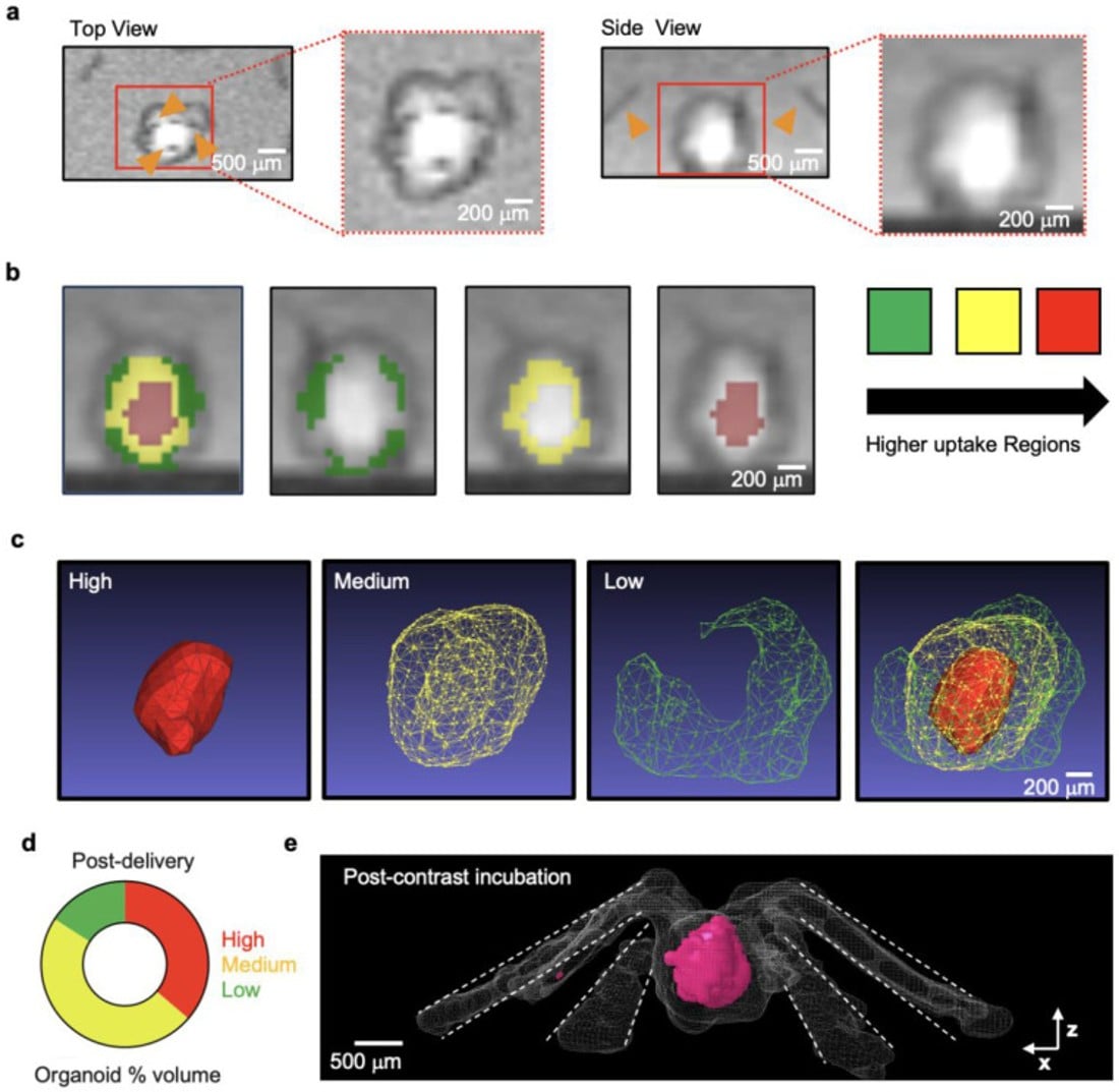
CTI also supports human-based testing models, which is an NIH research priority. Dr. Colin K. Franz, Associate Professor in the Departments of Physical Medicine & Rehabilitation and Neurology at NM Feinberg School of Medicine, is developing new approach methodologies (NAMs)—specifically, human stem cell–derived neural and neuromuscular organoids—to identify strategies that restore nerve and muscle function after injury or disease. A major limitation of NAMs is that cells found in the inner layers do not have constant access to nutrients in cell media; this restricts growth and cell survival. Dr. Franz’s team developed a 3D microfluidic delivery platform to integrate flexible, thread-like microchannels into growing organoids. These microchannels enable transport of nutrients to interior organoid layers.5 Using the MRI and CT instrumentation at CTI, Dr. Franz’s team generated 3D renderings of the organoid to demonstrate bidirectional exchange of solutes and successful diffusion of a contrast agent through the microchannels into the organoid (Figure 5).5 This study demonstrates the refinement of NAMs so that in vitro models more closely mimic human physiology, such as vascularization (blood flow through blood vessels), to empower disease modeling.
These studies highlight the importance of ORIP-funded research instrumentation to advance NIH priorities, such as chronic diseases and alternative testing models. Looking ahead, the ORIP-funded MRI scanner will play a pivotal role in supporting new research projects and emerging clinical translation efforts. ORIP’s investment in NM Feinberg School of Medicine’s imaging capabilities will allow the university to expand high-impact, translational biomedical imaging; foster new collaborations with external academic and industry researchers; and progress treatment strategies that improve health outcomes for patients with cancer or neurological diseases.
ORIP’s S10 programs support purchases of state-of-the-art, commercially available instruments to enhance research of NIH-funded investigators. S10 awards are made to domestic public and private institutions of higher education, as well as nonprofit domestic institutions, such as hospitals, health professional schools, and research organizations. Every instrument awarded by an S10 grant is to be used on a shared basis, which makes the programs cost efficient and beneficial to thousands of investigators in hundreds of institutions nationwide. For more information, please visit ORIP’s S10 Instrumentation Programs webpages.
References
1 Yan J, Kong LY, Hu J, et al. FGL2 as a multimodality regulator of tumor-mediated immune suppression and therapeutic target in gliomas. J Natl Cancer Inst. 2015;107(8):djv137. doi:10.1093/jnci/djv137.
2 Tripathi S, Najem H, Hurley L, et al. STING-induced blood-brain barrier opening combined with radiotherapy potentiates antitumor response in a high-grade glioma model. J Clin Invest. 2026;136(4):e198843. doi:10.1172/JCI198843..
3 Latha K, Yan J, Yang Y, et al. The role of fibrinogen-like protein 2 on immunosuppression and malignant progression in glioma. J Natl Cancer Inst. 2019;11(3):292–300. doi:10.1093/jnci/djy107..
4 Zannikou M, Duffy JT, Procissi D, et al. Bi-specific T cell-engaging antibody triggers protective immune memory and glioma microenvironment remodeling in immune-competent preclinical models. J Immunother Cancer. 2025;13(10):e011714. doi:10.1136/jitc-2025-011714..
5 Quezada MJ, Lee J, Lv Z, et al. Multiscale 3D microfluidic platform for intraorganoid delivery. Res Sq. 2026. doi:10.21203/rs.3.rs-8436544/v1.