


ORIP’s C06 Builds a Powerhouse for Preclinical Imaging and Irradiation Research at Wake Forest University School of Medicine
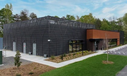
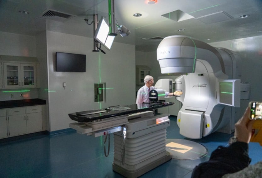
The Preclinical Imaging and Irradiation (PRIMIR) facility at Wake Forest University School of Medicine (WFUSM) is a state-of-the-art center that combines specialized imaging and radiation capabilities at a single location (Figure 1). PRIMIR is partnering with other leading research institutions—including Duke University, The University of North Carolina at Chapel Hill, University of Pittsburgh, and Columbia University—to conduct innovative research. Their collaborative pilot study with Duke University’s Radiation Countermeasures Center of Research Excellence (RadCCORE), for example, evaluated a novel radiomitigator compound using a state-of-the-art Varian TrueBeam™ 6 MV linear accelerator (Figure 2). Their compound helped promote rapid recovery of white blood cells after exposure to ionizing radiation, and the data will support countermeasure development against nuclear and radiation damage. Studies like this one highlight PRIMIR’s impact across the biomedical research landscape. In addition, Dr. George W. Schaaf, Assistant Professor in the Department of Radiation Oncology at WFUSM and Faculty Director for PRIMIR, noted that several collaborators are developing organoids and using artificial intelligence to support PRIMIR nonhuman primate (NHP) studies.
WFUSM has transformed its translational imaging and radiation research infrastructure with the opening of the PRIMIR facility, which was made possible by an ORIP grant (C06OD030099) awarded to the medical school. The PRIMIR facility eliminates the logistical hurdles and animal stress associated with transporting large animal models—including NHPs—across campuses for research studies. By consolidating imaging and radiation capabilities, WFUSM has created an environment where research can be conducted timely, efficiently, cohesively, and with significantly improved animal welfare outcomes. Dr. Schaaf emphasized that the PRIMIR facility supports NIH’s priority to strengthen research reproducibility. He elaborated, “Consolidation of the preclinical models with instrumentation on the same campus minimizes stress to our animals, enhancing animal welfare and the reproducibility of research results.”
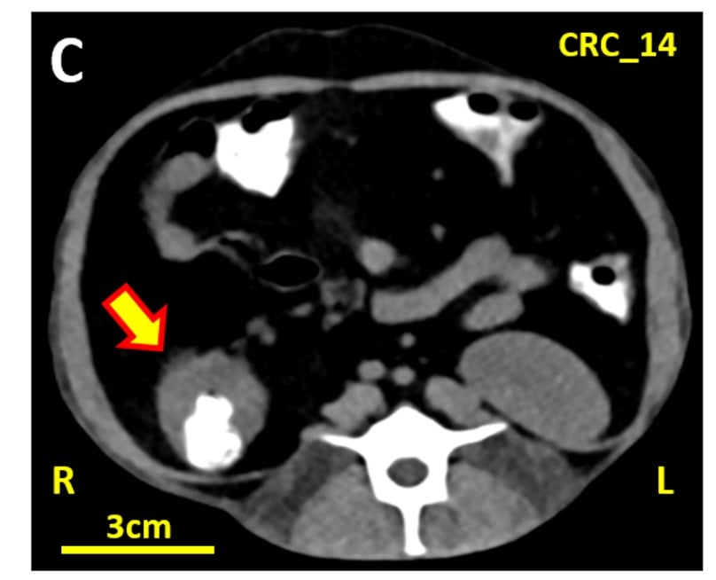
Since its grand opening in May 2025, the PRIMIR facility has provided researchers with access to two advanced imaging instruments—a positron emission tomography (PET)/computed tomography (CT) scanner and a 3 Tesla magnetic resonance imaging (MRI) scanner—to acquire cutting-edge images (Figure 3). Researchers also can utilize the third unique instrument—the Varian TrueBeam 6 MV linear accelerator—at PRIMIR for large-field radiation procedures and precision radiation (e.g., stereotactic body radiation therapy [SBRT]) research. The PRIMIR facility directly supports six major research programs spanning critical health challenges: Late Effects of Radiation, Alzheimer’s Disease, Aging, Substance Abuse, Neuro-Oncology, and Diabetes and Metabolic Disease.
Dr. Mark Baxter, Professor in the Department of Pathology, has a collaborative project with Dr. Carol A. Shively, Professor in the Department of Pathology, and Dr. Kiran K. Solingapuram Sai, Professor in the Department of Radiology and Director of the Translational Imaging Program (TIP), to study the underlying mechanisms of how diet composition affects cognition and brain aging. This study is supported by the National Institute on Aging (NIA) (RF1AG092490), and its results will guide dietary interventions that could help prevent neurodegenerative diseases. Dr. Solingapuram Sai also has funding from NIA (R01AG065839) to study the utility of novel small molecule–based probes with PET as an innovative imaging strategy. These probes target microtubules—a protein implicated in several pathologies, including Alzheimer’s disease.1 If successful, this tool will provide a noninvasive in vivo method for tracking neurodegeneration more easily and ensuring that therapeutic regimens are effective for patients. This innovative tool also is being explored in NHP models for radiation-induced brain injuries. Dr. Michael A. Nader, Professor in the Department of Translational Neuroscience, is using PRIMIR’s PET imaging to study brain changes in a preclinical model for cocaine use disorder. About 5 million people in the United States use cocaine,2 but no U.S. Food and Drug Administration–approved treatments exist for cocaine use disorder. With funding from the National Institute on Drug Abuse (R01DA017763), Dr. Nader’s research uses NHPs to parse out how variables, including sex and social rank, affect behavioral interventions and drug treatment outcomes. These studies have something in common—they depend on the instrumentation and model resources available at the PRIMIR facility, essential infrastructure supported by ORIP’s programs.
The PRIMIR facility also serves as the central hub for two national NIH-funded NHP resources: the Radiation Late Effects Cohort (U01AI150578) and the ORIP-funded Vervet Research Colony (P40OD010965). Dr. Schaaf and Dr. J. Mark Cline, Professor in the Department of Pathology, use PET/CT, MRI, and SBRT to support the Radiation Late Effects Cohort,3 which serves a highly active and collaborative network of researchers studying the aging-like multimorbidity phenotype induced by radiation exposure. This team also conducts proof-of-concept treatment studies for novel immunotherapies in NHP patients referred to WFUSM.4 Dr. Matthew J. Jorgensen, Professor in the Department of Pathology, is the principal investigator of the Vervet Research Colony, which serves a national collaborative network of more than 50 teams addressing research questions that attract the attention of multiple NIH institutes and centers.
Beyond immediate research applications, the PRIMIR facility is making strong contributions to workforce development in biomedical science. Dr. Dan Bourland, Professor in the Departments of Radiation Oncology and Biomedical Engineering and Physics, directs the WFUSM Radiation Physics Core. The linear accelerator is now the third specialized radiation device provided by the core, housed in the PRIMIR facility, offering unique research and educational opportunities for all trainees, such as the M.S. and Ph.D. graduate students enrolled in the university’s credentialed Biomedical Graduate Program in Medical Physics. In addition, the facility supports ORIP-funded T32 postdoctoral training (T32OD010957) and T35 summer veterinary student internships (T35OD010946), ensuring that the next generation of researchers gain hands-on experience with state-of-the-art imaging and radiation technologies and translational research methodologies.
Future expansion plans include installation of a cyclotron for short half-life radioactive isotope studies, which will further enhance research capabilities at WFUSM. The PRIMIR facility represents not just an infrastructure investment but a powerhouse for advancing research and health outcomes and training the next generation of biomedical scientists.
The PRIMIR facility was made possible through ORIP’s Research Facilities Construction Grant program (C06OD030099). ORIP’s Division of Construction and Instruments funds programs that support the construction, renovation, and modernization of research space by issuing notices of funding opportunities (NOFOs) when congressional appropriations are available. These programs aim to provide modernized physical infrastructure that meets the evolving engineering needs required to conduct cutting-edge NIH-funded biomedical research. These investments benefit nearly all NIH institutes and centers, advancing research across the full spectrum of biomedical sciences, from fundamental biology to clinical translational research.
References
1 Damuka N, Schaaf GW, Miller M, et al. Radiation-induced brain injury in non-human primates: A dual tracer PET study with [11C]MPC-6827 and [11C]PiB. Neuroimage Reports. 2025;5(1):100245. doi:10.1016/j.ynirp.2025.100245.
2 Key substance use and mental health indicators in the United States: Results from the 2023 National Survey on Drug Use and Health. Substance Abuse and Mental Health Services Administration. Published July 2024. Accessed April 17, 2026. https://www.samhsa.gov/data/sites/default/files/reports/rpt47095/National%20Report/National%20Report/2023-nsduh-annual-national.htm.
3 Olson JD, Schaaf GW, Bourland JD, Cline JM. The Wake Forest Nonhuman Primate Radiation Late Effects Cohort. Radiation Research. 2025;204(4):274–282. doi:10.1667/RADE-25-00063.1.
4 Deycmar S, Gomes B, Charo J, Ceppi M, Cline JM. Spontaneous, naturally occurring cancers in non-human primates as a translational model for cancer immunotherapy. Journal for Immunotherapy of Cancer. 2023;11(1):e005514. doi:10.1136/jitc-2022-005514.