


Micron-Scale Bioglass Confirmed with S10-Supported Optical Tweezers for Biomedical Research
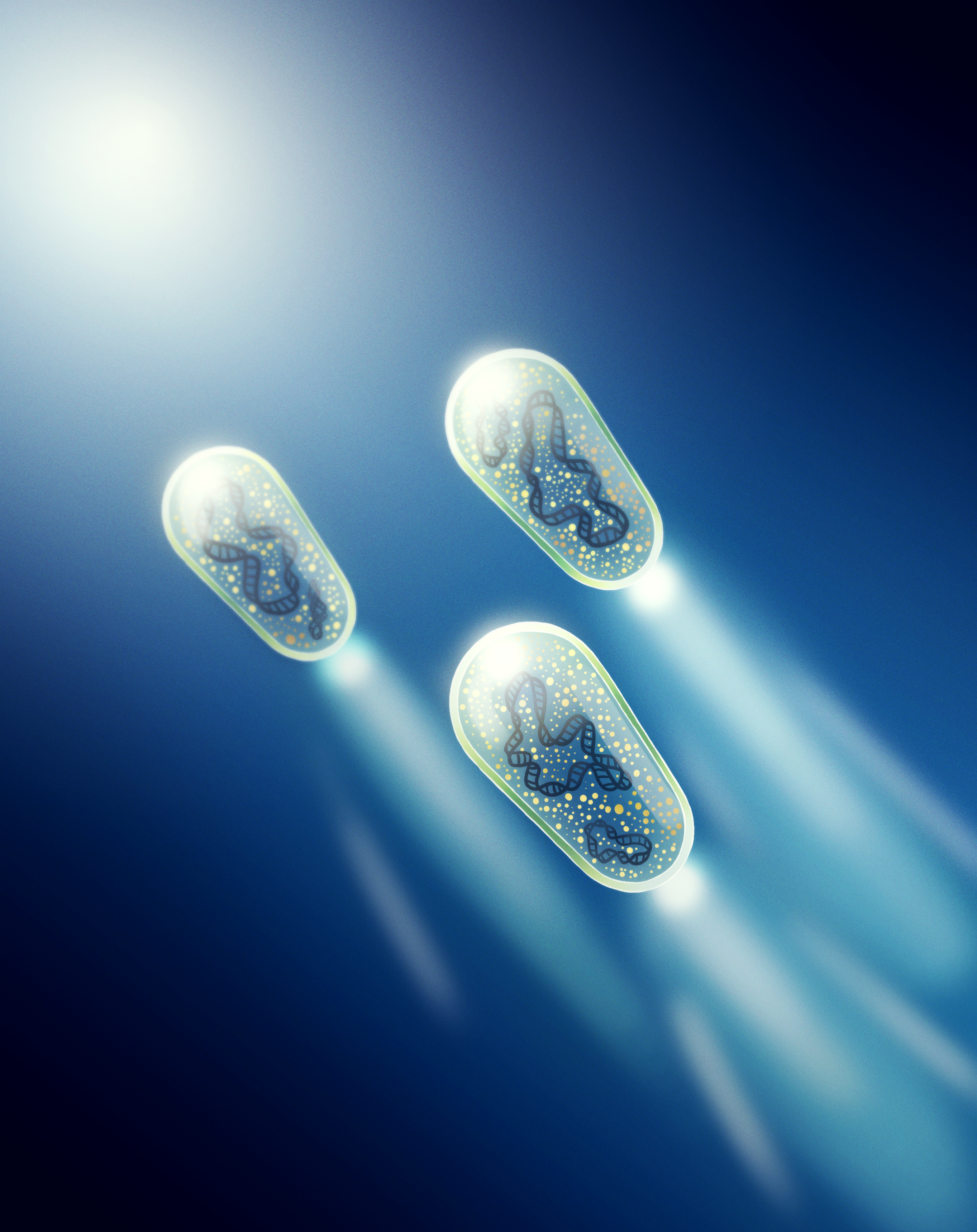
Biologically based glass (i.e., bioglass) has a longstanding history in health applications. Synthetic bioglass was developed in the late 1960s and has since been used in many ways.1 This silica-based, biodegradable material is less likely to be rejected by the body than many other materials used for medical implants. It also can bond with bone and has properties that aid in bone and tissue regeneration, and its predictable degradation makes it a useful tool for drug delivery. A team at the University of Rochester, headed by Dr. Lynn Sidor, Research Scientist II, and Dr. Anne Meyer, Associate Professor, is using a synthetic biology approach to produce tiny bioglass lenses (Figure 1) that can act as photonic devices. Their technique is inspired by similar processes that occur in nature. Technologies involving photonics—the use of light—have been widely applied in health care, including both diagnostics and treatment uses.2 These lenses are created at the micron length scale using Escherichia coli bacteria. The researchers can fine-tune these lenses, which enhances their potential application for biomedical research.
Tiny organisms and cells have been shown to exhibit lens-like properties for manipulating light, and other organisms create structures that can act as lenses.3 To create the bioglass lenses, the University of Rochester team used a type of sea sponge known as a glass sponge. These sponges harvest silica from the ocean to make glass elements that give them a stiff structure—essentially, these biological organisms create glass from a natural source, and the resulting material is called bioglass. This process is facilitated by a single enzyme, which the research team was able to insert into E. coli bacteria.3 In theory, this enzyme would encourage the bacteria to create glass lenses at small sizes that are difficult for today’s tools to achieve. "The bacterial cell is a really small object, and if you can get it to become this little photonic device, you’re working with sizes and tolerances that we don't have great ways to manufacture with other tools," explained Dr. Elio Abbondanzieri, Research Assistant Professor, Department of Biology, University of Rochester.
One challenge in the development of this bioglass is that the small size of the bacteria and thinness of the glass made confirming the presence and optical properties of the bioglass difficult. Dr. Abbondanzieri receives funding through ORIP’s S10 Shared Instrumentation Grant Programs (S10OD030296). With this funding, he obtained an instrument called “optical tweezers” to use in a variety of projects at the University of Rochester, including studying the bacteria-produced glass. Optical tweezers can trap small particles entirely through optical scattering forces. “Essentially, optical tweezers work as tractor beams, like from science fiction movies,” Dr. Abbondanzieri said. At that small scale, Brownian motion causes frequent movement, which can make seeing these tiny particles difficult. Optical tweezers create a “trap” to keep the cells in one place, nearly motionless. “It’s just frozen there, floating in the middle of water held by invisible forces that you can’t see, and suddenly you can see everything, every little motion that it makes down to the nanometer level, thanks to all of the light that’s going through it,” Dr. Abbondanzieri explained.
With the bacteria trapped by the optical tweezers, the motion that still occurs can be measured precisely using video tracking and characteristic patterns of light scattering. Few instruments are sensitive enough to assess the difference in light scattering between bacterial cells with several nanometers of glass on the outside and those without. The optical tweezers enabled the team to confirm that the optical properties of the bacteria had indeed changed, proving that these cells had produced the bioglass coating as intended. When optical tweezers are used to trap two materials with different indices of refraction, differences between variations in the size of the particles versus variations in the material are clear. This measurement was verified using polystyrene and silica beads, and the team was able to conduct a similar test on bacteria with and without bioglass encapsulation. “Now we could separate the size of the bacteria versus how tightly they’re trapped,” Dr. Abbondanzieri explained. “When we plot that in two dimensions, these two types of bacteria lie on diagonal lines that are separated from each other. Now we actually had the ability to say with confidence and statistics that this glass encapsulation really changes how the light interacts with these bacteria.”
The bioglass produced by the bacteria had several exciting properties, hinting at the broad potential these tiny lenses have in real-world applications. "One thing that they do, which was really striking, is that the cells encapsulated in the glass were much better at focusing light," Dr. Abbondanzieri said. Dr. Abbondanzieri's team found that a beam of light cast through the bioglass-encapsulated E. coli cells would exit the cell as a photonic nanojet—a beam of light that is even smaller than the limits of the light's wavelength would suggest. This ability provides significant promise for using bioglass cells to replace regular glass in technological applications far beyond what existing technologies can offer. For example, the current microlenses used in phone cameras are larger than E. coli cells and could be replaced by these lenses made from natural materials. “People are always trying to get more megapixels on their camera, meaning the detectors have to be broken into smaller and smaller squares," Dr. Abbondanzieri commented. “Bacteria, being naturally a very small shape, are poised to fit into those devices quite well."
The team is exploring ways to use bioglass cells for other nanophotonics purposes. Many of these approaches are relevant to health care applications. For example, these lenses can be used to direct light to specific locations, a technique currently used in photodynamic therapy for treatment of tumors.4 Additionally, these tiny cells can be used as biosensors that identify differences in light properties caused by changes within an organism, such as shifts in blood composition that signal early signs of disease.5 "We look at it as pointing the way for other applications," Dr. Abbondanzieri said. "This technique gives you a range to play with in terms of sizes and shapes of cells other than E. coli that you could put these genes into." Dr. Abbondanzieri expressed gratitude to ORIP for its support of the optical tweezers purchased through the S10 grant and the many scientific advancements it has contributed to. "This is an instrument that lets people do types of experiments that they just couldn't do any other way," he emphasized. "It's opening up new possibilities, and I'm excited to see what else people will be doing with this in the future."
ORIP's S10 programs support purchases of state-of-the-art, commercially available instruments to enhance research of NIH-funded investigators. S10 awards are made to domestic public and private institutions of higher education, as well as nonprofit domestic institutions, such as hospitals, health professional schools, and research organizations. Every instrument funded by an S10 grant is to be shared with other institutions, which makes the programs cost efficient and beneficial to thousands of investigators in hundreds of institutions nationwide. For more information, please visit ORIP's S10 Instrumentation Programs webpage.
References
1 Cannio M, Bellucci D, Roether JA, et al. Bioactive glass applications: A literature review of human clinical trials. Materials (Basel). 2021 Sep 20;14(18):5440. doi:10.3390/ma14185440.
2 Sarbadhikary P, George BP, Abrahamse H. Paradigm shift in future biophotonics for imaging and therapy: Miniature living lasers to cellular scale optoelectronics. Theranostics. 2022 Oct 17;12(17):7335–7350. doi:10.7150/thno.75905.
3 Sidor LM, Beaulieu MM, Rasskazov I, et al. Engineered bacteria that self-assemble bioglass polysilicate coatings display enhanced light focusing. Proc. Natl. Acad. Sci. U.S.A. 2024;121(51):e2409335121. doi:10.1073/pnas.2409335121.
4 National Cancer Institute. Photodynamic therapy to treat cancer. https://www.cancer.gov/about-cancer/treatment/types/photodynamic-therapy.
5 Li Y, Liu X, Xu X, et al. Red-blood-cell waveguide as a living biosensor and micromotor. Adv. Funct. Mater. 2019;29:1905568. doi:10.1002/adfm.201905568.