The WormGUIDES Atlas: A Window into the Mysteries of Neurodevelopment in Caenorhabditis elegans
More than 30 years after the adult nervous system and cell lineage of the roundworm Caenorhabditis elegans were first mapped,1 that map of neuron connectivity (i.e., the connectome) still enables scientists to better understand diverse neurobiological mechanisms. Today, C. elegans remains a widely used model for neuroscience research because of its short life cycle, transparent body, and homology to human genes expressed in neurodevelopment. C. elegans offers an ideal system for visualizing neurodevelopment and for modeling neurodegenerative diseases, such as Alzheimer’s disease and autism spectrum disorder (ASD). This model, however, has limitations with regard to how well researchers can visualize neurodevelopment over time and the absence of the necessary single-cell resolution.

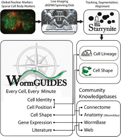
Supported by the National Institutes of Health (NIH) Office of Research Infrastructure Programs (ORIP), Dr. Daniel Colón-Ramos at Yale University, Dr. Zhirong Bao at the Sloan Kettering Institute, Dr. Hari Shroff at the NIH, and Dr. William Mohler at the University of Connecticut established a consortium called WormGUIDES (A Resource for Global Understanding in Dynamic Embryonic Systems), which provides a system-level resource on the developing embryonic nervous system of C. elegans at the single-cell level. By integrating light-sheet microscopy, imaging, and computational approaches, consortium investigators developed the WormGUIDES Atlas, an interactive resource that provides dynamic information in four dimensions (4D) on the behaviors of individual cells as they coordinately assemble into a functional nervous system in the C. elegans embryo (Figure 1). The consortium also developed a mobile version of the WormGUIDES Atlas for Android and iOS platforms.2
WormGUIDES principal investigator and project lead, Dr. Colón-Ramos, explained that during development, hundreds of neurons coordinately assemble into the circuits that underpin behavior. The WormGUIDES Atlas provides a unique window into this process, documenting and enabling discovery-driven exploration of how the networks of the entire nervous system form. Because these processes are conserved in evolution, understanding the organizing principles in a worm brain will illuminate scientist’s understanding of human brain organization and development and, most importantly, how these processes go awry and cause disease.
“The fundamental biological question that researchers do not understand is how organs form in general. In a complex organ, like the brain, many different cells precisely wire to form circuits. The brain allows the organism to perceive its environment, to learn from it, and to respond accordingly,” said Dr. Colón-Ramos. He explained that the goal of the WormGUIDES consortium is to use C. elegans to understand these developmental neurological processes. The consortium resulted in the creation of a system that tracks single cells through time during development. During the past 6 to 7 years, WormGUIDES researchers have been developing new technologies for capturing early neurodevelopmental events and new methods for analyzing and sharing systems-level data with the community (wormguides.org). These efforts have resulted in new resources for investigators, including WormGUIDES Data, WormGUIDES Ultrastructure, and a database of neuron-specific marker genes and expression patterns.
WormGUIDES has facilitated several important research findings. For example, in 2019, WormGUIDES research teams expanded knowledge on morphogenesis of the nervous system by visualizing neurons in a C. elegans sensory organ (called the amphid) as it underwent a collective dendrite extension to form the amphid’s sensory system.3 WormGUIDES researchers developed an integrated protocol for examining neurodevelopment in C. elegans embryos, which was made available to other investigators.4
WormGUIDES Atlas shows particular promise as a resource for studying the mechanisms of human neurodevelopmental diseases. According to Dr. Colón-Ramos, “Many diseases remain mysterious to us because we only understand the emergent property—such as the phenomenology of the disease, the symptoms—but we don’t understand its underpinning. One disease that could benefit from the tools and resources developed is ASD.” Dr. Colón-Ramos added that an important goal of the WormGUIDES consortium is to better understand the processes involved in normal neurodevelopment work and how these processes change in disease. The consortium is laying the necessary groundwork for understanding these processes, which Dr. Colón-Ramos anticipates will provide insight into the mechanisms of diseases that have remained a mystery to neuroscientists for far too long.
Looking to the future of WormGUIDES, Dr. Colón-Ramos described plans to develop orthogonal approaches complementary to what has been achieved, such as time-series observations via electron microscopy and X-ray studies to understand nanoscale precision in embryo circuit connectivity during development. The WormGUIDES consortium of biologists, computer scientists, and microscopists continues to conduct bimonthly meetings, collaborate on publications, and train and mentor junior investigators.
References
1 White JG, Southgate E, Thomson JN, Brenner S. The structure of the nervous system of the nematode Caenorhabditis elegans. Philos Trans R Soc Lond B Biol Sci. 1986;314(1165):1–340.
2 Santella A, Catena R, Kovacevic I, et al. WormGUIDES: An interactive single-cell developmental atlas and tool for collaborative multidimensional data exploration. BMC Bioinformatics. 2015;16:189.
3 Fan L, Kovacevic I, Heiman MG, Bao Z. A multicellular rosette-mediated collective dendrite extension. Elife. 2019;8:e38065.
4 Duncan LH, Moyle MW, Shao L, et al. Isotropic light-sheet microscopy and automated cell lineage analyses to catalogue Caenorhabditis elegans embryogenesis with subcellular resolution. J Vis Exp. 2019;148:e59533.


