Super-Resolution Imaging: Beating the Boundaries of Light
Substance use disorders and neuropsychiatric conditions are brain diseases characterized by morphological and functional adaptations in neurons and neural circuits. Traditionally, neuroscientists use conventional confocal or electron microscopy to characterize how abused substances remodel sites of synaptic communication. However, confocal microscopy is hindered by diffraction limits of light waves and cannot resolve structures below a spatial resolution of about 250 nm. Although electron microscopy has a much higher resolution (2 nm) than light microscopy, obtaining and analyzing neural structures, such as synapses and dendritic spines, in three-dimensions are often not feasible due to experimental constraints and high costs. Even though there are some tools available to examine morphological and functional changes in the brain following alcohol and drug exposure, inherent limitations of these tools have curbed our progress of understanding how drugs of abuse affect synapses and other cellular sub-compartments.
Recent technological and computational advances, termed ‟super-resolution” imaging, allow to see beyond the diffraction limit and into a biological relevant range for small substructures, like dendritic spine necks. Spine necks are crucial neuronal sub-compartments with a diameter of about 150 nm, below the detection limit of confocal microscopy. The narrow necks act as a spatial filter, isolating spino-somatic voltage signals and limiting biochemical diffusion from spines into parent dendritic shafts.1,2 This structure is critical for enhancing the fidelity of signaling between neurons. Moreover, changes in spine neck morphology may relate to aberrant neuronal communication resulting from alcohol and drug use/misuse. For example, a collaborative study from the NIH’s National Institute of Alcohol Abuse and Alcoholism (NIAAA)-funded Neurobiology of Adolescent Drinking in Adulthood (NADIA) Consortium revealed that adolescent alcohol exposure reduced the diameter and volume of dendritic spine necks in dentate gyrus granule neurons.3,4 In this study, the U.S. Food and Drug Administration (FDA)-approved cholinesterase inhibitor, donepezil (Aricept), reversed the effect of adolescent alcohol exposure on spine neck morphology. Despite these intriguing findings, the molecular mechanisms driving the morphological adaptations remain unknown.

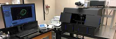
The NIH’s Office of Research Infrastructure Programs (ORIP), through its Shared Instrumentation Program (S10), supports grant awards for the acquisition of the state-of-the art instruments that enable and enhance research of NIH-supported investigators. Dr. Patrick Mulholland, an Associate Professor in the Departments of Neuroscience and Psychiatry & Behavioral Sciences at the Medical University of South Carolina (MUSC), received an S10 award to support the purchase of a confocal microscopy system with a super-resolution detector. Installed in June 2017, the Carl Zeiss LSM 880 confocal microscope with an Airyscan super-resolution detector provides a two-fold increase in axial and lateral resolution and an eight-fold improvement in the signal-to-noise ratio. This versatile confocal system also is equipped with the fast Airyscan module that allows high-speed signal acquisition in living specimens while maintaining super-resolution structural information.
The addition of this microscope helped to establish a Shared Confocal Core Facility (SCCF)5 for biomedical researchers at MUSC, under the supervision of Dr. Mulholland. The SCCF supports neuroscientists studying addiction and neuropsychiatric conditions, providing faculty, postdoctoral fellows, and graduate students with training and access to state-of-the-art imaging equipment. In addition, the SCCF gives users access to high-end workstations running 3D image analysis and deconvolution software that can render and analyze large-scale images (100+ GB) collected with the super-resolution Airyscan detector.
A team of researchers at MUSC led by Dr. Mulholland employs the microscope to examine morphological adaptations in cellular sub-compartments induced by abused substances and neuropsychiatric conditions. Since its installation, Dr. Mulholland’s group has demonstrated that the super-resolution detector enhances the ability to detect dendritic spines and provides high-resolution images of their morphology, including spine heads and necks.6 In addition, studies performed for the NADIA Consortium using the Airyscan detector have revealed sex differences in the morphology of dendritic spines in the lateral septum, a brain region critical for social behavior, mood, and stress responsivity (P. Mulholland, L. Spear, D. Werner, and E. Varlinskaya, unpublished observation). Finally, the microscope is used for more refined analyses of neuroadaptations in cortical regions in relation to alcohol dependence and relapse drinking, a major focus of the NIAAA-supported P50 grant awarded to the Charleston Alcohol Research Center at MUSC.
This technological advance in the resolution of fluorescent imaging provides exciting opportunities to characterize how alcohol and other abused drugs shape dendritic spine morphology and how they modify elements that organize spine sub-compartments. Indeed, studies using super-resolution imaging approaches have identified molecular mechanisms that coordinate the structural organization of dendritic spines.7 Through its super-resolution imaging capabilities and enhanced signal-to-noise ratio, the super-resolution microscope and image analysis software available to MUSC’s investigators through the SCCF will enhance the understanding of mechanisms and processes that drive substance use disorders, as well as neuropsychiatric conditions, such as depression, anxiety, and Autism Spectrum Disorder.
References
1Jayant K, et al. Targeted intracellular voltage recordings from dendritic spines using quantum-dot-coated nanopipettes. Nature Nanotechnology 2017;12:335-342, doi:10.1038/nnano.2016.268.
2Tonnesen J, Katona G, Rozsa B, Nagerl U V. Spine neck plasticity regulates compartmentalization of synapses. Nature Neuroscience 2014;17:678-685, doi:10.1038/nn.3682.
3Mulholland P J, et al. Donepezil reverses dendritic spine morphology adaptations and Fmr1 epigenetic modifications in hippocampus of adult rats after adolescent alcohol exposure. Alcoholism: Clinical and Experimental Research 2018;42:706-717, doi:10.1111/acer.13599.
4Dovey D. Alzheimer’s drug repairs binge drinking brain damage in mice, 2018, https://www.newsweek.com/binge-drinking-teen-drinking-brain-damage-alcohol-809532.
5Shared Confocal Core Facility, https://medicine.musc.edu/departments/neuroscience/research/mulholland
6McGuier NS, Uys JD, Mulholland PJ. In: Neural Mechanisms of Addiction (Ed. M. Torregrossa) Elsevier, 2018.
7MacGillavry HD and Hoogenraad CC. The internal architecture of dendritic spines revealed by super-resolution imaging: What did we learn so far? Exp Cell Res 2015;335:180-186, doi:10.1016/j.yexcr.2015.02.024.


