S10-funded Microscopes Propel UT Southwestern Facilities to the Forefront of Research
Imaging plays a pivotal role in deciphering the molecular pathways of human physiology and pathophysiology. At The University of Texas Southwestern Medical Center (UT Southwestern), four instruments funded through ORIP’s S10 Shared Instrumentation Programs have contributed significantly to this role. These instruments, which were awarded between fiscal years 2015 and 2020, are a scanning electron microscope (S10OD020103), a laser scanning confocal microscope (S10OD021684), a transmission electron microscope (S10OD021685), and a spinning disk confocal microscope (S10OD028630). Located in the Live Cell Imaging and Electron Microscopy core facilities, these instruments have been used by more than 50 research groups from approximately 20 departments campus wide.

Dr. Katherine Luby-Phelps, Professor in the Department of Cell Biology at UT Southwestern, serves as Director of the two facilities in which the funded instruments are housed. With her extensive expertise in electron and light microscopy, Dr. Luby-Phelps oversees technical staff and contributes to a broad array of scientific projects. She explained that the S10 grants have been crucial for ensuring the continuation of imaging services for the facilities’ users.
“The S10 program has really been the way in which we have been able to replace aging equipment or, in some cases, the way in which we’re able to acquire new technologies,” Dr. Luby-Phelps stated. “Without it, I don’t know where we’d be in terms of being able to provide [our] services.”
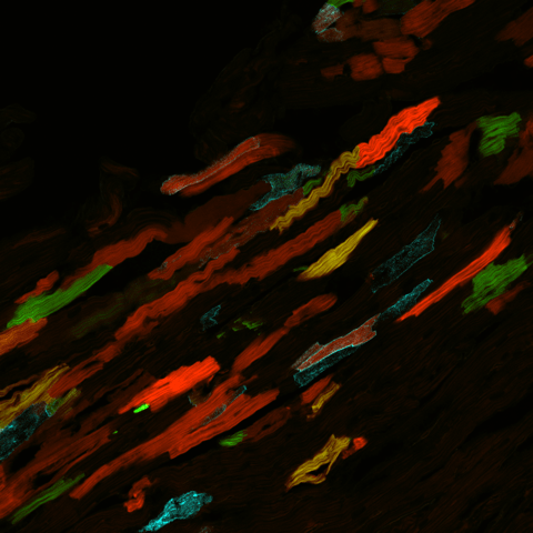
Dr. Luby-Phelps explained that the different microscopes can be applied to various areas of research to yield insight on biological structures at different spatial scales. Scanning electron microscopy, for example, is essential for research investigating the surface structures of biological samples. Transmission electron microscopy, in contrast, allows researchers to examine inner ultrastructural features of cells at high resolution. Confocal microscopes can be used for immunofluorescent imaging of cellular components (Figure 1). The spinning disk confocal microscope offers advanced capabilities for live-cell imaging.

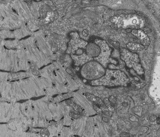
These instruments have supported numerous research projects advancing understanding of the origin and treatment of human diseases and have led to many high-profile publications. For example, the S10-funded transmission electron microscope has been used by UT Southwestern investigators to study how the loss of a specific nuclear envelope protein leads to profound structural defects in the skeletal muscle of mouse models for Emery Dreifuss muscular dystrophy (Figure 2).1
Another application of the instrumentation is stent angioplasty, which is used widely in the treatment of both congenital and adult heart disease. Conventional stents have excellent short term results but are plagued with long-term thrombosis and restenosis. To address these issues, Tre Welch, Ph.D., Assistant Professor in the UT Southwestern Department of Cardiovascular and Thoracic Surgery, and his team are designing and optimizing expandable, bioresorbable stents constructed from poly(L-lactic acid) (PLLA) fibers and testing them in animal models.2 Scanning electron microscopy using the S10-funded instrument is an essential tool for examining the torsion of the PLLA fibers and changes in the structural integrity of the stent fibers over time (Figure 3).

Dr. Luby-Phelps explained that most of the facilities’ users are affiliated with UT Southwestern and funded by the NIH, although outside users also have access to the facilities’ services. The Electron Microscopy Core Facility, in particular, serves a large base of external users, including investigators from institutions located outside the United States. Dr. Luby-Phelps also emphasized that the instruments are used continuously as valuable learning tools for students and postdoctoral researchers; these services are crucial for the science education and professional training of these users.
Reflecting on her experiences as both an S10 Shared Instrumentation Program awardee (i.e., she has successfully applied for program funding) and reviewer (i.e., she served on peer review panels assessing the scientific merit of applications requesting microscopes), Dr. Luby-Phelps underscored the value of the program for the research community. “I think the S10 program is the most useful program that NIH provides,” she stated. “It’s essential for keeping the infrastructure of our research institutions at the forefront, keeping them modern and up to date. … Being involved with the S10 program on both sides of the fence has been a really rewarding experience, and I just can’t say enough good things about the program.”
References
1 Ramirez-Martinez A, et al. The nuclear envelope protein Net39 is essential for muscle nuclear integrity and chromatin organization. Nat Commun. 2021;12: 690. doi: 10.1038/s41467-021-20987-x.
2 Wright J, et al. Bioresorbable stent to manage congenital heart defects in children. Materialia (Oxf). 2021;16:101078. doi: 10.1016/j.mtla.2021.101078.


