Microscope Funded by S10 Program Advances Biological Research at the University of Delaware
Located within the heart of the Delaware Biotechnology Institute Bio-Imaging Center, the Zeiss LSM 880 inverted laser scanning confocal microscope is a valuable asset for researchers at the University of Delaware (UD). The microscope, which was secured through a grant awarded in 2018 through ORIP’s S10 Instrumentation Programs (S10OD016361), is capable of such techniques as simple and multicolor imaging, spectral unmixing, and super-resolution imaging.
Powered by a laser beam that scans biological samples, the LSM 880 can provide unique insights into biological structures. Dr. Jeffrey Caplan, director of the Bio-Imaging Center, emphasized that the LSM 880’s power lies in its remarkable flexibility.
“Because we have such a wide user base at the University of Delaware, we don’t focus on just one particular area like cancer or neuroscience. We’re a research institution that has really a wide range of scientists,” Dr. Caplan stated. “That’s what’s exciting about that microscope—it’s agnostic to the biological samples that you put on there.”

The LSM 880 serves approximately 20 individual research groups per year, with an average of 45 to 60 investigators. Of those users, approximately 80% to 85% are associated with UD. The remaining users represent outside academic institutions and private industry. Dr. Caplan explained that the Bio-Imaging Center offers a critical combination of the technology and expertise needed to perform the imaging techniques across disciplines and biological questions. Since its installation, the instrument has yielded 41 publications.
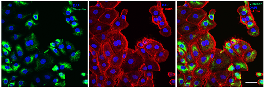
At UD, many professors have used the LSM 880 for new discoveries in their research. One of the instrument’s most active users is Dr. Xinqiao Jia, who synthesizes hydrogels that create 3D microenvironments that enable an improved understanding of cell growth and tissue formation (Figure 1).1,2 Dr. Emily Day has used the instrument to characterize nanoparticles that contain microRNA.3 Dr. John Slater has achieved photo-ablation in hydrogels, mimicking biological structures (Figure 2).4 Dr. Caplan, who is funded in part by the National Institute of General Medical Sciences, also uses the instrument for his research on organelle and molecular movements within cells.5
Recent upgrades—including the addition of an Airyscan detector and a multiphoton laser—resulted in a twofold increase in the instrument’s resolution. Dr. Caplan emphasized that these upgrades have provided new capabilities for users who previously were working at the edge of the instrument’s resolution. These upgrades represented a combination of internal and external funds, including an Institutional Development Award (IDeA) Networks of Biomedical Research Excellence grant (P20GM103446). IDeA is a congressionally mandated program that builds research capacity in states—including Delaware—that historically have had low levels of NIH funding.
Several years after its installation, the LSM 880 remains an invaluable resource at UD. Dr. Caplan underscored that access to shared instrumentation is vital for both senior investigators and their graduate students, who represent the next generation of scientists.
“I just looked at the calendar, and [the microscope] is booked all day, 7 days a week,” Dr. Caplan recounted. “It’s still a heavily used microscope, and I suspect it will be for years to come.”
References
1 Zerdoum AB, Fowler EW, Jia X. Induction of fibrogenic phenotype in human mesenchymal stem cells by connective tissue growth factor in a hydrogel model of soft connective tissue. ACS Biomater Sci Eng. 2019;5(9):4531–4541. doi: 10.1021/acsbiomaterials.9b00425.
2 Liu S, et al. Cellular interactions with hydrogel microfibers synthesized via interfacial tetrazine ligation. Biomaterials. 2018;180:24–35. doi: 10.1016/j.biomaterials.2018.06.042.
3 Kapadia CH, Ioele SA, Day ES. Layer-by-layer assembled PLGA nanoparticles carrying miR-34a cargo inhibit the proliferation and cell cycle progression of triple-negative breast cancer cells. J Biomed Mater Res A. 2020;108(3):601–613. doi: 10.1002/jbm.a.36840
4 Banda OA, Sabanayagam CR, Slater JH. Reference-free traction force microscopy platform fabricated via two-photon laser scanning lithography enables facile measurement of cell-generated forces. ACS Appl Mater Interfaces. 2019;11(20):18233–18241. doi: 10.1021/acsami.9b04362.
5 Kumar AS, et al. Stromule extension along microtubules coordinated with actin-mediated anchoring guides perinuclear chloroplast movement during innate immunity. Elife. 2018;7:e23625. doi: 10.7554/eLife.23625.


