Immunological Dysfunction in Long COVID Patients Characterized Using ORIP S10-Funded Equipment

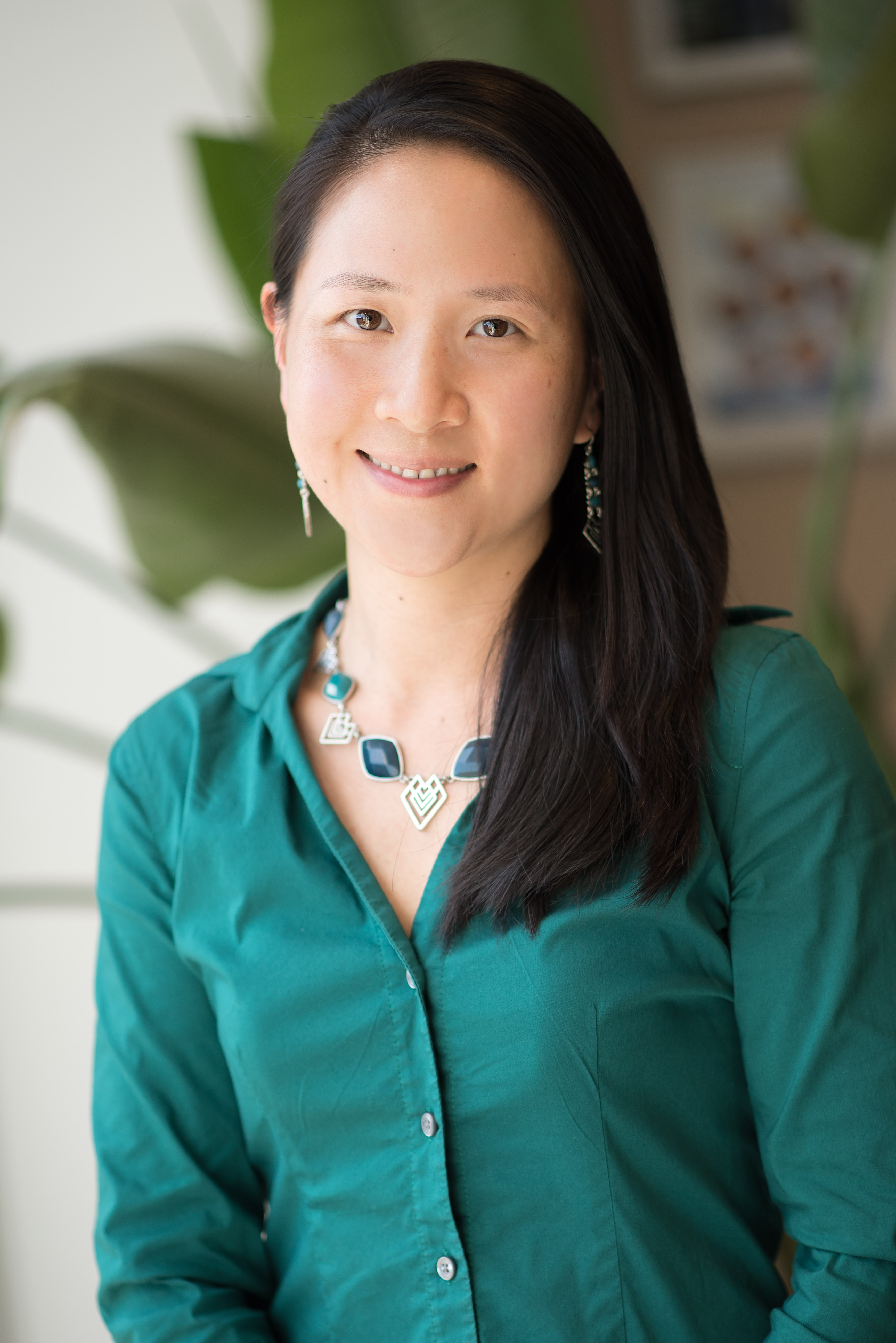
According to a 2024 survey by the National Center for Health Statistics, approximately 10% of adults who have had a COVID-19 infection report having long COVID.1 Long COVID is characterized by a range of distressing symptoms that can last for weeks, months, or even years after an initial case of COVID-19 has been resolved. Common symptoms of long COVID include fatigue, brain fog, joint or muscle pain, shortness of breath, and loss of taste or smell, and researchers are just beginning to dissect out the potential mechanisms underlying this condition. After studying the effects of acute SARS-CoV-2 infection on the immune system, Dr. Nadia Roan (Figure 1), a Senior Investigator at the Gladstone Institute of Virology, turned her attention to long COVID. Using a technique called cytometry by time-of-flight (CyTOF) and instruments supported by an S10 Shared Instrumentation Grant Program award from ORIP, Dr. Roan’s laboratory and her colleagues at the University of California, San Francisco (UCSF) identified significant changes in immune cells from the blood of individuals experiencing long COVID. The study sheds light on possible underlying causes of long COVID and uncovers biological changes that might serve as biomarkers of long COVID or targets for therapies to cure this debilitating condition.
Dr. Roan is a Professor of Urology at UCSF and a virologist with expertise in microbial pathogenesis and immunology. Before the COVID-19 pandemic, her research focused on the role that T cells play during HIV infection. T cells are white blood cells that help mount an immune response to infectious pathogens. Cytotoxic T cells (or CD8+ T cells) destroy infected cells. Helper T cells (or CD4+ T cells) emit molecular signals that direct other immune cells to fight infections. “T cells are really important effectors against many different kinds of viruses, including HIV,” explained Dr. Roan. “One of the earliest observations made during the initial stages of the pandemic was that the people who had the most severe COVID also had the most profound depletion of T cells. It really highlighted the notion that T cells likely play a key role in immunity against SARS-CoV‑2, which was not surprising given the essential role of T cells in defense against viruses.”
To better understand the state of the immune system in patients with long COVID, Dr. Roan’s group analyzed blood cells from patients with long COVID and compared the results to fully recovered patients. The blood samples were taken from the Long-term Impact of Infection with Novel Coronavirus (LIINC) cohort, which was established at the beginning of the COVID-19 pandemic and has collected longitudinal specimens from more than 1,000 individuals so far. “We picked individuals who got COVID and then had continuous symptoms throughout the first initial eight months,” noted Dr. Roan, “versus those who got symptomatic COVID-19 (which was confirmed by PCR test) but where the symptoms went away after acute infection and did not return.” Importantly, at the 8month time point when samples analyzed in this study were collected, the individuals had not been vaccinated or reinfected, both of which can alter features of immune cells.
To maximize the information captured from such valuable samples, Dr. Roan’s group used CyTOF, a variation of flow cytometry in which proteins of interest are labeled and detected using heavy metal ions rather than fluorescent tags. When compared with standard flow cytometry, CyTOF enables quantitation of more proteins on and inside a cell. “I think it’s a very powerful technology,” observed Dr. Roan. “We used the CyTOF to characterize the features of both total T cells from these individuals, as well as those specifically recognizing SARS-CoV-2.” The experiments were conducted by members of Dr. Roan’s laboratory using a Helios CyTOF instrument at the UCSF Parnassus Flow Cytometry CoLab. This instrument was purchased by UCSF with support from an S10 award (S10OD018040) provided by ORIP in 2014. To validate the CyTOF results, the blood samples were analyzed using flow cytometry, as well as single-cell RNA sequencing. The flow cytometry experiments were performed on a Fortessa X-20 flow cytometer that was purchased by the Gladstone Institutes with support from an S10 award (S10RR028962) provided by ORIP in 2011.

Dr. Roan and her team observed no difference in the total numbers of SARS-CoV-2-specific T cells between long COVID and recovered patients (Figure 2). However, the distributions of certain subsets of T cells were altered. Notably, higher levels of multiple chemokine receptors (which direct T cells to migrate into various tissues) were found on the surface of CD4+ T cells in long COVID patients than in the same cells in recovered patients. SARS-CoV-2-specific CD8+ T cells from patients with long COVID also expressed higher levels of exhaustion markers, suggesting cellular dysfunction, which can arise after chronic exposure to a persistent virus. The group also discovered that patients with long COVID showed miscoordination between their T-cell-mediated response and humoral (i.e., antibody) response to SARS-CoV-2. SARS-CoV-2-specific antibody levels correlated with SARS-CoV-2-specific T-cell levels (CD4+ and CD8+) in recovered patients but not in patients with long COVID. Long COVID patients who had the highest levels of SARS-CoV-2-specific T cells also had almost undetectable levels of SARS-CoV-2-specific antibodies. The study was published in the January 2024 issue of Nature Immunology.2
Dr. Roan described the significance of these results: “The antibody arm of immunity typically coordinates together with the T-cell arm of immunity. I like to think of it as an orchestra that is working together in synchrony. But when the coordination is off, and you have these individuals, for example, who have a high antibody response but almost no detectable T-cell response (and vice versa), that can lead to problems.” The varying T-cell characteristics observed between recovered patients and those with long COVID are consistent with the hypothesis of a tissue reservoir of SARS-CoV-2-infected cells that cannot be cleared in long COVID patients. “Perhaps the CD4+ cells are migrating into tissues because there is inflammation there due to persistent virus. The preferential expression of exhaustion markers among SARS-CoV-2-specific CD8+ T cells in long COVID patients is also consistent with viral persistence. When virus cannot be cleared, the T cells that recognize them can become exhausted. This previously has been observed in the context of HIV, a classic example of a persistent virus.”
Dr. Roan reflected on how her background in HIV research enabled the COVID-19 studies: “The reason we were able to quickly implement CyTOF-based analysis of T cells at the beginning of the COVID-19 pandemic was that we had already developed a variety of CyTOF-based tools for our HIV work.” Her group had previously used both S10-supported instruments (in combination with bioinformatic approaches) to investigate HIV persistence. “Both the CyTOF instrument and the flow cytometer were crucial in our studies to characterize the phenotypic features of HIV reservoir cells in antiretroviral therapy–suppressed people with HIV,” emphasized Dr. Roan.
Dr. Roan plans to continue working with the LIINC cohort to study long COVID. Her group intends to move beyond the blood and investigate immune cells that are present in various tissue compartments. She also noted that interventional trials—including those testing the effects of SARS-CoV-2 antivirals—have been initiated within the LIINC cohort and pointed out that samples from before and after treatment would be especially informative. Dr. Roan added that her group, working together with the laboratories of Dr. Melanie Ott and Dr. Jorge Palop at the Gladstone Institutes, has developed a mouse model for SARS-CoV-2 infection that can be used to further explore COVID-19 interventions. The team already has shown that mild infection in these mice causes simultaneous behavioral and immunological changes that extend into the post-acute stages of infection. “I think this model is really powerful,” remarked Dr. Roan, “because we can test things like a prolonged Paxlovid treatment course or other antivirals and see whether any of these treatments alleviate these behavioral and immunological abnormalities. I think pairing the knowledge gleaned from these mouse studies together with clinical studies in people will be quite informative.”
For more information, please visit ORIP’s S10 Instrumentation Programs webpages.
References
1 National Center for Health Statistics. U.S. Census Bureau, Household Pulse Survey, 2022–2024. Long COVID. https://www.cdc.gov/nchs/covid19/pulse/long-covid.htm
2 Yin K, Peluso MJ, Luo X., et al. Long COVID manifests with T cell dysregulation, inflammation and an uncoordinated adaptive immune response to SARS-CoV-2. Nat Immunol. 2024;25(2):218–225. doi:10.1038/s41590-023-01724-6.


