“Going with the Flow”—Biological Cell Sorting
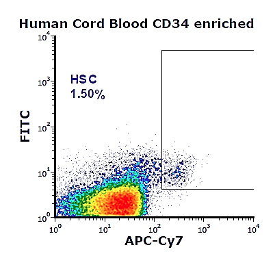
Many of the scientific discoveries require an understanding of specialized biological processes at the cellular level. Scientists rely on the use of light microscopes to visualize cells and other organisms invisible to the human eye. To segregate cell populations from a heterogenous mixture and to quantitatively analyze cells from a variety of biological specimens, many scientists employ flow cytometry. Flow cytometry is a laser-based technology to identify and measure specific parameters of cell populations in a high-throughput manner1, allowing the simultaneous analysis of cell-surface and intracellular characteristics of thousands of cells per second in real time. This technology is made possible via a fluidic system where individual cells labeled with fluorescent markers (fluorochromes), flow in a liquid stream.This stream of cells pass through multiple laser beams that excite the fluorochromes; their signals are then quantified by an electronic detector. Such sorting method, referred to as Fluorescent Activated Cell Sorting (FACS), isolates sub-populations of cells, allowing their functional characterization and cell-to-cell interaction studies.2
Why is it important to sort cells? Most diseases can be linked to the dysfunction of cell populations in a larger complex biological environment. Isolating cells from clinical samples by a cell sorter is an ideal method to purify specific cells of interest; for example, from the bone marrow, circulating blood stream, digestive system tissues, tumors, or other bodily specimens. Cell sorting and the downstream analysis can assist in the diagnosis of a variety of disorders and provide data regarding cellular mechanisms that may eventually lead to the development of new treatments. FACS has redefined the way scientists analyze cells and advanced biomedical research in immunology, cancer biology, drug screening, and also contributed to clinical diagnostics.1,2 Broad utility of the FACS technology has created a big demand for cell sorters.
In the last 6 years, ORIP's S10 Instrumentation Grant Program awarded about 75 cell sorters and cell analyzers to about 60 institutions nationwide. This includes a 2015 award to Columbia University Medical Center to acquire a state-of-the-art Becton Dickinson [BD] Biosciences Influx™ cell sorter.3 The instrument was installed in the Columbia Center for Translational Immunology (CCTI) flow cytometry core facility as a shared instrument.4
CCTI serves several departments within Columbia, supporting research on stem cells, neurobiology, and cancer. Experienced staff at CCTI, including Dr. Siu-Hong Ho (Assistant Professor of Medical Sciences, Manager of CCTI), ensures the effective use of the instrument. The Influx cell sorter has an advanced "jet-in-air" technology: when cells are injected into the machine, their speed in the fluid remains constant, reducing stress on cells caused by the injection process. The machine offers up to 10 lasers and has a flexible platform for flow cytometry that can adapt to the end-user's research requirements. Dr. Ho adds "The sorting speed and purity of post-sort survival cells is incomparable to other sorters, especially for fragile and time-sensitive cells", such as as activated T cells and neurons.

Under the leadership of Dr. Hans-Willem Snoeck (Professor of Medicine, Department of Microbiology and Immunology), the Influx cell sorter has been used by researchers across the institution and in collaboration with other scientists for investigating specialized cells of the immune system -T and B cells - that are involved in responses to transplantation, in mechanisms of autoimmune disorders, and genetic-based diseases.
The laboratory of Dr. Snoeck conducts multidisciplinary research to gain better understanding of immunology-based diseases. The goal is to improve bone marrow transplantation and gene therapy methods that target hematopoietic stem cells (HSCs). HSCs are specialized stem cells that bring about all other blood cells through cell differentiation. The Influx cell sorter is being used in studies that advance understanding of (1) the mechanisms that induce a state of immunological tolerance, (2) the role of the cell machinery mitochondria in HSC function, and (3) how to isolate specialized cells from tissues to elucidate their function and homeostasis.
The use of the Influx cell sorter has contributed to substantial high-impact scientific findings. These include the discovery by Dr. Snoeck's laboratory that the mitochondria play an important role in maintaining HSCs that have the potential to differentiate into lymphoid cells.5 This finding holds an important clinical relevance by providing new approaches to bias HSCs differentiation into the desired cells after their transplantation into a patient. In addition, HSCs are candidates for the treatment of various disorders, such as sickle cell disease.
Further highlighting the use of the Influx cell sorter, the research team of Dr. Megan Sykes, also from Columbia University, developed an assay to identify donor T cells that are tolerogenic in recipient kidney and bone marrow transplant patients.6 Tolerogenic T cells that can inhibit chronic rejection of donor organs in patients may result in increased survivability for recipients. Since the installation of the instrument, the research on the BD Influx cytometer awarded by ORIP has yielded dozens of high-impact publications and initiated many ongoing projects producing groundbreaking data.
References
1 Adan A, Alizada G, Kiraz Y, Baran Y, Nalbant A. Flow cytometry: basic principles and applications. Critical Reviews in Biotechnology. 2017; 37(2):163-176.
2 Lanier, L. Just the FACS. The Journal of Immunology. 2014;193(5):2043-2044.
3 BD Influx Cell Sorter, https://www.bdbiosciences.com/us/instruments/research/cell-sorters/bd-influx/m/744777/overview
4 CCTI Core Facility, http://www.cumc.columbia.edu/ccti/research/cores/flow-cytometry-core
5 Luchsinger, L., de Almeida, M, Corrigan, D, Mumau, M, Snoeck, H.W. Mitofusin 2 maintains haematopoietic stem cells with extensive lymphoid potential. Nature. 2016;529(7587):528-531.
6 Morris H, DeWolf S, Robins H, Sprangers B, LoCascio SA, Shonts BA, et al. Tracking donor-reactive T cells: Evidence for clonal deletion in tolerant kidney transplant patients. Science Translational Medicine. 2015;7(272): 272ra10. doi:10.1126/scitranslmed.3010760.


