


S10 Instrument Contributed to the Development of Patented Silent MRI Technique for Functional Neuroimaging
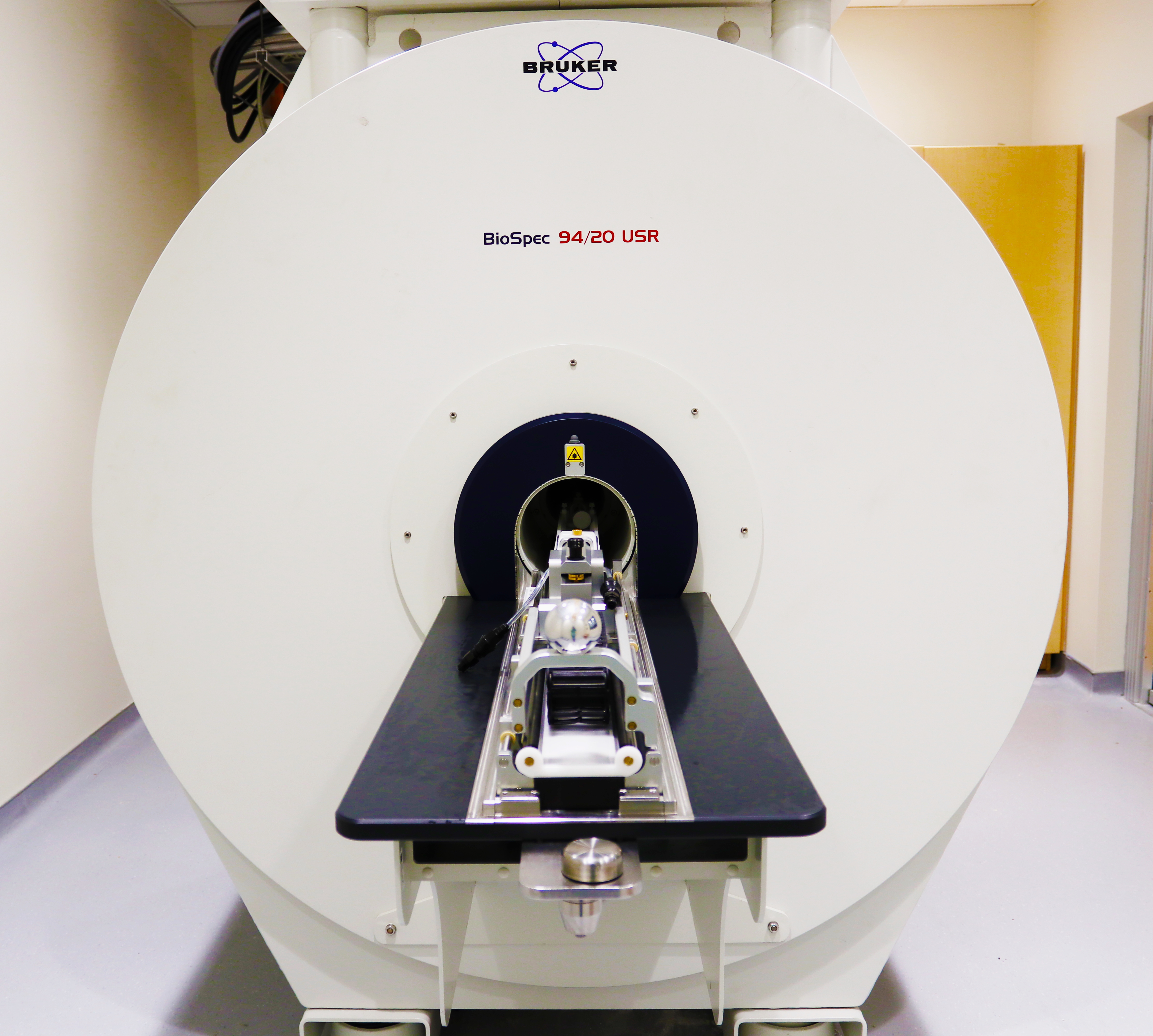
The relationship between behavior and brain-wide activity is difficult to study in animal models. This is because the technology frequently used to view activity changes in the brain—magnetic resonance imaging (MRI)—generates high acoustic noise, which can stress the animal and is sensitive to movement, creating artifacts in the imaging. At The University of North Carolina (UNC) School of Medicine, Dr. Yen-Yu Ian Shih, Professor and Vice Chair for Research, Department of Neurology; Director, Center for Animal MRI; and Associate Director, Biomedical Research Imaging Center, and his team have used their 9.4T small animal MRI scanner (Figure 1), funded by the UNC School of Medicine Office of Research and an S10 Shared Instrumentation Grant from ORIP (S10OD026796), to develop a silent functional MRI (fMRI) technique to advance the imaging of small animals and other sensitive study subjects.
Dr. Shih explained that one of the main challenges in animal MRI is that the noise of the machine panics the animals into moving, which produces unwanted signals in the images and complicates determination of brain functional status. “The level of sound we are generating in typical MRI scans is equivalent to a lawn mower,” he said. “Now imagine placing a little mouse on the ground and driving a lawn mower right next to it.” Although animals can become accustomed to the sound, this takes significant time. Anesthetizing the animals is a common option, but it introduces complications, as brain dynamics under anesthesia may differ from those in awake states, making it harder to achieve a clean study of naturalistic activities and behaviors.
To address these challenges, Dr. Shih and his team developed a novel MRI data sampling strategy that minimizes acoustic noise during scans. This new method, termed Steady-state On-the-Ramp Detection of INduction-decay with Oversampling (SORDINO),1 also overcomes a number of limitations in current standard fMRI techniques. SORDINO significantly reduces motion and magnetic field susceptibility artifacts while providing robust sensitivity and specificity in detecting brain activity. This method opens new possibilities for imaging whole-brain function in awake, behaving mice, which was previously very challenging, if not impossible. SORDINO—named for a term that instructs a musician to mute an instrument—eliminates the acoustic noise of the MRI machine, allowing fMRI studies of the mouse brain during active behaviors, such as seeing, reaching for, and eating food pellets, or engaging in social interactions. By correlating the fMRI data with observed behaviors, researchers can gain a more holistic view of the brain and its communication dynamics.
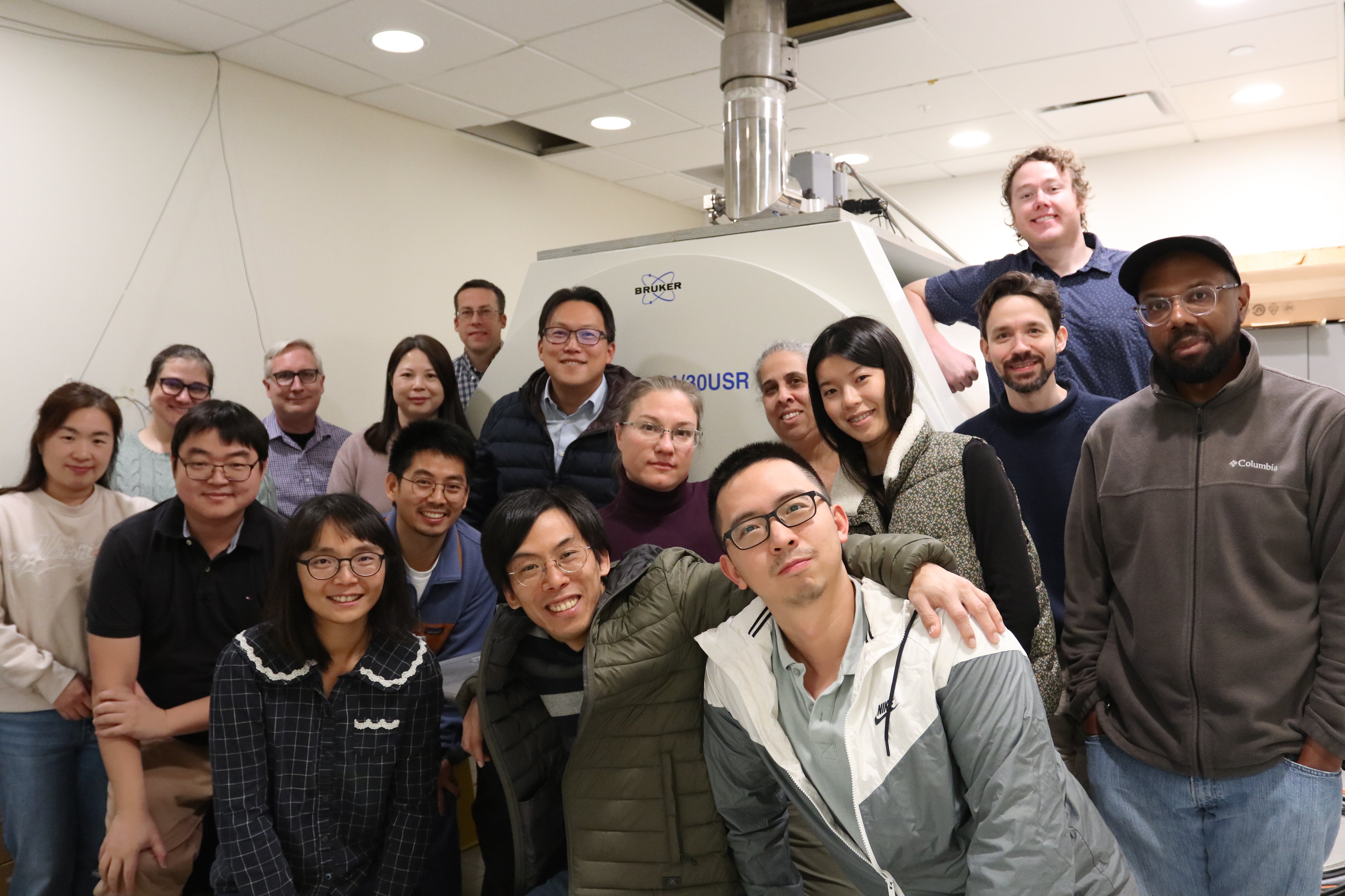
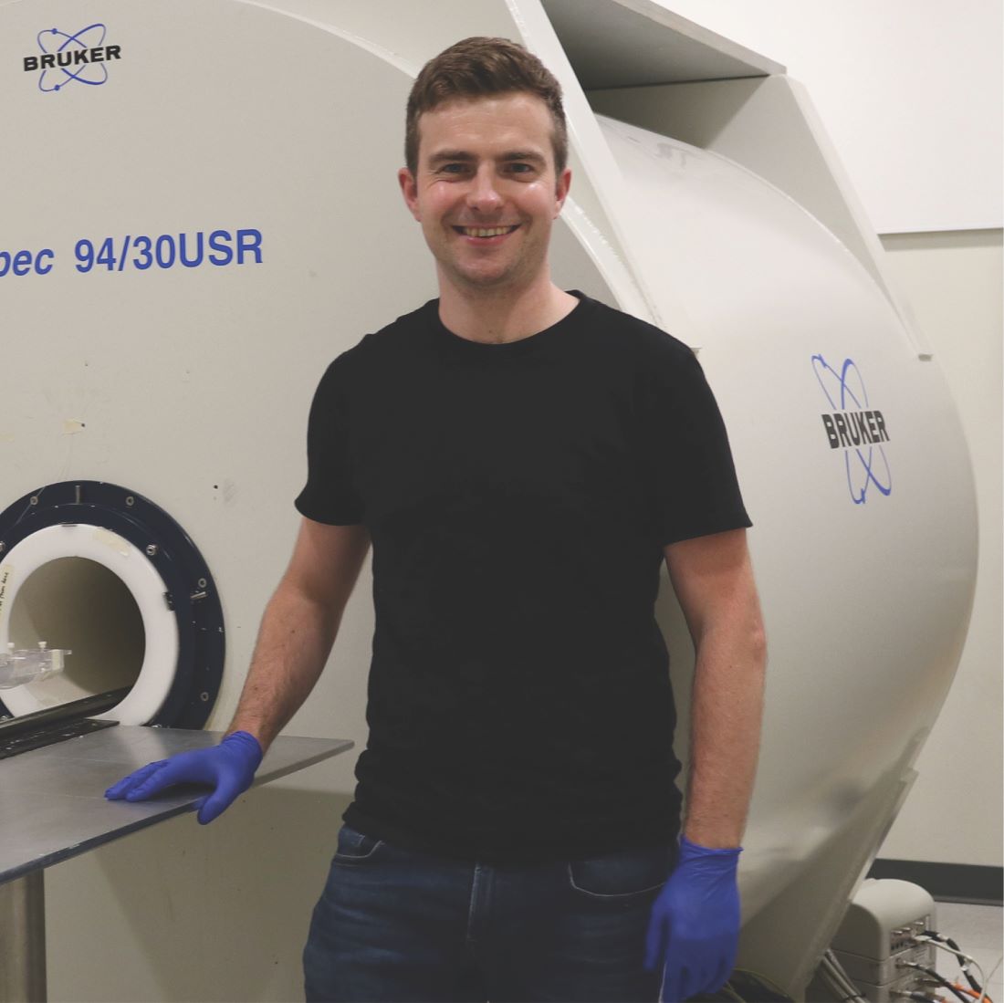
Dr. Shih and his team (Figures 2 and 3) were granted a U.S. patent2 for the originality and innovation of their work in 2024. They plan to disseminate the technique to the research community in hopes of expanding and improving biomedical research at a broad scale. “What’s remarkable about this method is that it’s primarily a software solution,” Dr. Shih explained. “It can be implemented on any type of MRI system and adapted for various applications.” One of Dr. Shih’s goals is to extend the application of the SORDINO technique to human MRI. Although the acoustic noise and motion artifacts in MRI are usually tolerable for healthy adult human subjects, they present challenges for studying certain patient populations and pose a major barrier to performing research on the developing human brain in infants, where both noise and motion impact imaging quality.
Dr. Shih emphasized the importance of developing technologies to drive meaningful scientific progress. His team’s innovation represents a great example that builds on previous discoveries to deliver impactful improvements to a widely used technology. As such, this innovation is the perfect marriage of Dr. Shih’s research interests in better understanding brain circuitry and optimizing technology to advance research. He directs the Center for Animal MRI at the UNC Biomedical Research Imaging Center, a research unit and core facility that supports a variety of projects for investigators interested in using MRI systems. Dr. Shih has overseen a transformative increase in research capacity since obtaining the ORIP-funded 9.4T small animal MRI scanner. “This S10 makes us a hub for advanced animal MRI research,” Dr. Shih said. “With the most up-to-date equipment—perhaps the newest electronics available in the field—we can deliver very high-quality images to our users and support their innovations.” Many researchers come to the Center to conduct experiments in areas other than neuroscience, so the instrument is used for a variety of research studies by investigators from across the nation. Dr. Shih and his team are committed to developing and refining protocols to meet the unique needs of their collaborators, which will foster advancements across a broad range of scientific communities.
Dr. Shih credited ORIP’s support for making such progress possible. “An instrument of this scale is not easy for a single research lab to house and manage,” Dr. Shih said. “The S10 program provides an invaluable platform for institutions like ours.” He emphasized that this support plays a crucial role for state institutions, where securing funding for large-scale instruments can be especially challenging. “This program truly levels the playing field and allows a broader base of researchers to access cutting-edge technologies,” he said.
ORIP’s S10 programs support purchases of state-of-the-art, commercially available instruments to enhance research of NIH-funded investigators. S10 awards are made to domestic public and private institutions of higher education, as well as nonprofit domestic institutions, such as hospitals, health professional schools, and research organizations. Every instrument awarded by an S10 grant is to be used on a shared basis, which makes the programs cost efficient and beneficial to thousands of investigators in hundreds of institutions nationwide. For more information, please visit ORIP’s S10 Instrumentation Programs webpages.
References
1MacKinnon MJ, Song S, Chao THH, et al. SORDINO for silent, sensitive, specific, and artifact-resisting fMRI in awake behaving mice. bioRxiv 2025.03.10.642406. doi:10.1101/2025.03.10.642406.
2Shih Y, MacKinnon MJ, Ma Y, Chang W. Methods and systems for functional magnetic resonance imaging with a zero echo time pulse-sequence. United States patent US11957444B2, filed August 8, 2022, and issued April 16, 2024.